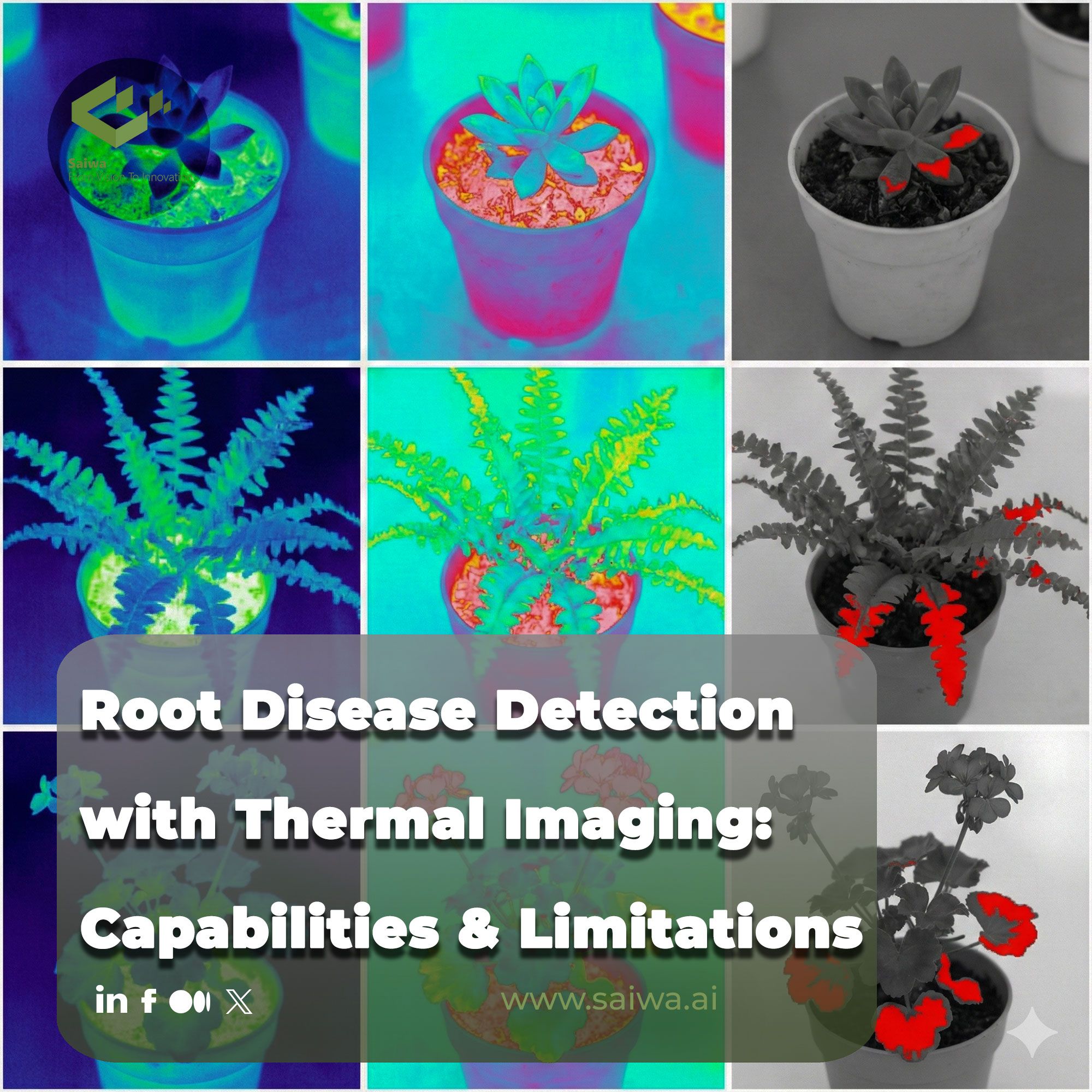
Root Disease Detection with Thermal Imaging: Opportunities & Limitations
Explore how thermal infrared imaging and machine learning enable early, non-destructive detection of root diseases in crops before visible symptoms appear.
Written by Amirhossein
Reviewed by Boshra
Introduction
Before we start we would like to have a quick introduction at thermal imaging concept. Nowadays, thermal imaging has emerged as a powerful sensing tool in precision agriculture. It offers new possibilities for non-destructive and continuous monitoring of plant health. By capturing temperature variations associated with physiological processes, thermal infrared (TIR) imaging provides indirect but yet sensitive indicators of plant stress. In this section we introduce the fundamental principles of infrared thermography, highlights the importance of early detection of root diseases from both agricultural and economic perspectives, and discusses the limitations of conventional plant disease diagnostic methods. Together, these topics establish the scientific and practical motivation for adopting thermal imaging technologies in modern crop monitoring systems and its application in root desease detection as our main interest in this article.
Thermal Infrared Imaging and Thermography
TIR imaging, also known as infrared thermography, is a non-contact sensing technique that measures the thermal radiation emitted by objects in the infrared region of the electromagnetic spectrum. In plant phenotyping and disease monitoring, TIR imaging is primarily used to estimate surface temperature variations that are associated with physiological processes such as transpiration and stomatal regulation. Infrared thermography operates mainly in the long-wave infrared range (approximately 8–14 µm), where vegetation exhibits high emissivity and stable radiative behavior, allowing accurate surface temperature estimation under controlled conditions .
In agricultural applications, thermography enables the continuous and non-destructive observation of plant responses to both biotic and abiotic stresses. Temperature differences detected at the canopy or leaf level are frequently associated with changes in water status, metabolic activity, or vascular disruption caused by disease progression. As we will see further in this section, this technique has been increasingly integrated into precision agriculture frameworks due to its capability to capture early stress indicators before visual symptoms appear .
Early Root Disease Detection: Economic and Agricultural Impact
Before going further we should notice that root diseases pose a major challenge to crop productivity because they interfere directly with water and nutrient uptake, that often leads to reduced growth, yield loss, and plant mortality. There are several studies emphasize that root and crown diseases frequently remain undetected until aboveground symptoms become visible, at which point physiological damage is already advanced and often irreversible . Early detection is therefore critical for timely intervention and disease management.
Infrared thermography has been proposed as a promising approach for early disease detection because physiological alterations caused by root pathogens can induce measurable thermal responses in aerial plant parts. These temperature changes are associated with disruptions in transpiration and vascular function, which can be detected prior to the appearance of chlorosis, wilting, or necrosis . If we can identify stress at an early stage, it supports improved decision-making in crop protection and resource management.
Limitations of Traditional Plant Disease Diagnostics
For plant disease detection, going to conventional methods rely largely on visual inspection, destructive sampling, and laboratory-based techniques such as pathogen isolation or molecular assays. While these methods can provide accurate identification of pathogens, they are often time-consuming, labor-intensive, and unsuitable for large-scale or continuous monitoring that are the real cases that fast and accurate detections matter a lot. On the other hand, visual assessments are inherently subjective and typically detect disease only after significant physiological damage has occurred.
Destructive sampling methods that we face in traditional methods disrupt plant integrity and cannot be applied repeatedly to the same specimen, limiting their usefulness in longitudinal studies or real-time monitoring systems . In contrast, non-destructive sensing techniques such as infrared thermography that we are going to study more in this article, offer the potential for rapid screening of plant health without physical contact, enabling repeated measurements and early stress detection .

Root Diseases and Their Thermal Signatures
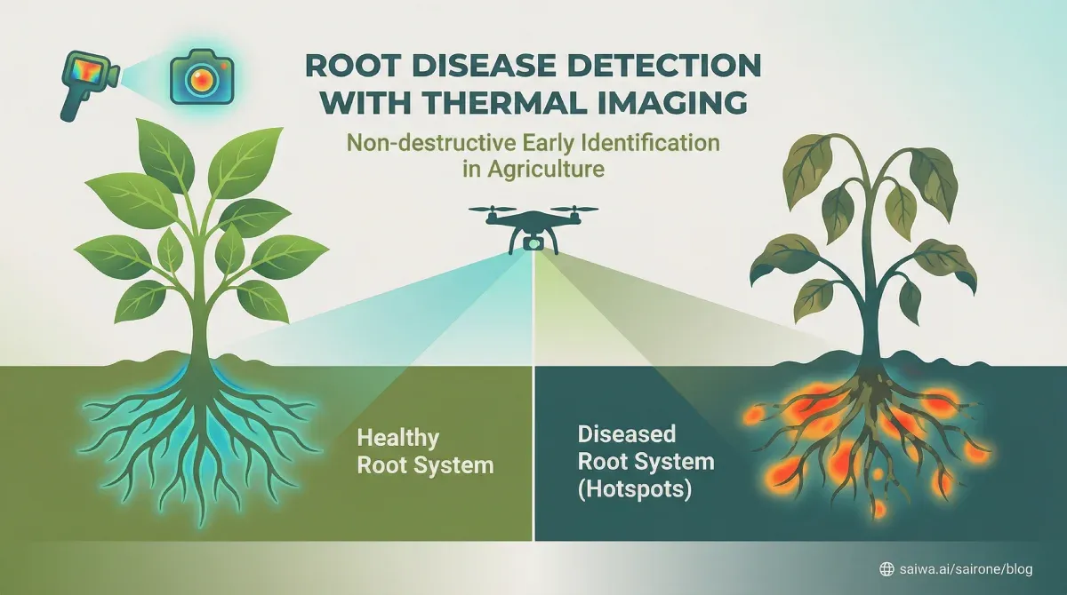
After this introcution, let's have a look at Root disease issue. Root diseases represent a major class of biotic stresses that originate belowground yet exert significant effects on aboveground plant physiology. Soil-borne pathogens disrupt root function, water uptake, and vascular transport, leading to measurable changes in plant thermal behavior. In this section we focus on common root pathogens and parasitic plants that explains how root infections alter physiological processes related to transpiration and temperature regulation, and explores the relationship between root stress, canopy temperature, and leaf surface temperature (LST). Emphasis is also placed on the ability of thermal imaging to detect early stress responses before visible disease symptoms emerge, highlighting the relevance of thermal signatures as indirect indicators of root health.
Common Soil-Borne Pathogens: Fusarium, Rhizoctonia, Sclerotinia, and Orobanche
Firsrt we should know soil-borne pathogens are responsible for a wide range of root and crown diseases that significantly affect agricultural crops. Among these, Fusarium species are frequently studied due to their widespread distribution and severe impact on plant vascular systems. Fusarium root and crown rot have been shown to impair water transport and induce physiological stress that manifests in altered thermal patterns at the canopy level .
While Fusarium is explicitly examined in experimental thermal imaging studies, other soil-borne pathogens such as Rhizoctonia and Sclerotinia are previously discussed more generally in the context of plant disease monitoring and stress physiology . These pathogens similarly disrupt root function and water balance, which can theoretically lead to detectable temperature anomalies in aboveground tissues. Parasitic plants such as Orobanche affect host plants by extracting water and nutrients from the root system, potentially inducing stress responses comparable to those caused by pathogenic infections .
Root Diseases Affecting Plant Physiology and Temperature
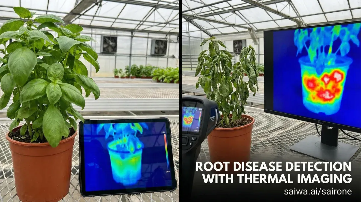
Root diseases interfere with fundamental physiological processes by damaging root tissues and vascular connections. This disruption limits water uptake and transport to aerial parts of the plant, leading to reduced transpiration and altered stomatal behavior . As transpiration plays a central role in leaf cooling, any reduction in transpiration rate can result in elevated leaf and canopy temperatures.
Experimental observations demonstrate that plants infected with Fusarium root and crown rot exhibit higher mean leaf surface temperatures compared to healthy plants under similar environmental conditions . These thermal differences reflect the physiological stress imposed by root pathogens and provide a measurable indicator of disease presence.
The Connection Between Root Stress, Canopy Temperature, and Leaf Surface Temperature
Canopy temperature and leaf surface temperature (LST) are closely linked to plant water status and stomatal conductance. When root systems are compromised by disease, the resulting water stress leads to partial or complete stomatal closure, reducing transpiration and increasing surface temperature . Thermal imaging captures these temperature variations, enabling indirect assessment of root health through aboveground measurements.
In the literature, several studies highlight that temperature-based indicators derived from thermal images correlate with physiological stress responses rather than specific pathogen presence . As a result, thermal signatures must be interpreted within the broader context of plant–environment interactions and supporting diagnostic information.
Detecting Early Stress Before Visible Symptoms Emerge
One of the primary advantages of infrared thermography is its ability to detect physiological stress prior to the development of visible disease symptoms. In controlled experiments, temperature differences between healthy and infected plants were observed before morphological changes such as wilting or discoloration became apparent . This early detection capability is particularly valuable for root diseases, which often progress unnoticed below the soil surface.
We showed that thermal imaging provides a temporal advantage over traditional diagnostic approaches by revealing stress-related temperature anomalies at an early stage of infection so let's have a deeper look at TIR.
Thermal Infrared Imaging Technology
Up to know we undersood that TIR imaging technology provides the technical foundation for capturing and interpreting plant temperature information in agricultural applications. As our next step, understanding how thermal cameras measure infrared radiation and convert it into surface temperature values is essential for accurate data acquisition and analysis. This section introduces the operating principles of thermal imaging sensors, including wavelength ranges, spatial and thermal resolution considerations, and the advantages of non-destructive testing (NDT) for plant health monitoring. Additionally, key parameters such as surface emissivity and its standardization for vegetation are discussed, as they play a critical role in ensuring reliable and comparable temperature measurements across different experimental conditions.
Thermal Cameras and Sensors
Thermal cameras detect infrared radiation emitted by objects and convert this radiation into temperature values based on physical laws governing thermal emission. The measured radiance depends on object temperature, emissivity, and environmental factors such as atmospheric absorption . In plant applications, accurate temperature measurement requires consideration of surface emissivity and sensor calibration.
Infrared thermography systems generate two-dimensional temperature maps that represent spatial variations across leaves or canopies. These thermal images serve as the basis for quantitative analysis and feature extraction in plant stress and disease detection studies .
To be more specific, most agricultural thermal imaging systems operate in the long-wave infrared (LWIR) range, typically between 8 and 14 or 15 µm, where vegetation exhibits high and stable emissivity . Sensor resolution and thermal sensitivity are critical parameters that influence the ability to detect subtle temperature differences associated with early disease stress.
Higher spatial resolution allows more detailed characterization of temperature heterogeneity across plant surfaces, while high thermal resolution improves sensitivity to small temperature variations . These sensor characteristics will directly affect the performance of downstream image analysis and classification tasks.
Non-Destructive Testing
It is interesting to know that infrared thermography is classified as a NDT technique because it enables plant assessment without physical contact or tissue damage. This property allows one repeating measurements over time, supporting longitudinal monitoring of disease progression and plant response to environmental conditions . This Non-destructive thermal monitoring preserves plant integrity and is particularly suitable for greenhouse experiments and controlled studies where repeated observations of the same plants are required .
Emissivity in Plant Measurements: Standardization
Another key parameter is emissivity which is a critical parameter in thermal imaging, representing the efficiency with which a surface emits infrared radiation. We know that vegetation is commonly assumed to have a high emissivity value, typically around 0.95, which allows reliable temperature estimation when this parameter is properly configured in thermal cameras . Additionally, standardizing emissivity settings is essential for obtaining comparable temperature measurements across different experiments and environmental conditions. Incorrect emissivity assumptions can lead to systematic temperature errors and misinterpretation of thermal data .
Active vs. Passive Thermography Methods
Active and passive methods are widly used in plant disease detection. It depends on whether an external energy source is applied during image acquisition or not. These two approaches differ fundamentally in how thermal information is generated and interpreted, and each presents distinct advantages and limitations in agricultural applications. But in plant pathology and stress monitoring, passive thermography is more commonly employed, as it captures naturally emitted thermal radiation associated with physiological processes such as transpiration and stomatal regulation . Active thermography, by contrast, involves controlled thermal excitation and has been discussed mainly in methodological contexts, with limited direct application in plant disease detection studies. Understanding the principles, capabilities, and constraints of both methods is essential for selecting appropriate thermal imaging strategies, particularly when targeting subtle stress responses induced by root diseases . So, let's have a deeper look at these two types in the sequel.
Passive Thermography
As we mentioned earlier, passive infrared thermography is the most commonly applied approach in plant disease detection. In this mode, thermal radiation that naturally emitted by plant surfaces is recorded without applying any external heat source. Temperature variations captured through passive thermography reflect physiological processes such as transpiration rate, stomatal conductance, and water status of plants . It also allows continuous and non-destructive monitoring under greenhouse and field conditions. But measurements are highly sensitive to environmental factors including air temperature, humidity, and wind conditions.
Active Thermography
On the other hand, active thermography involves the application of an external energy source to induce thermal responses in plant tissues, followed by monitoring heat dissipation patterns. This approach has been mainly discussed in the context of material inspection and is less frequently applied in agricultural plant disease studies. We see limited references that suggest active methods may help enhance thermal contrast by stimulating physiological responses, but practical implementation for root disease detection remains underexplored in plant pathology literature .
Comparison
Most plant disease detection studies, particularly those focusing on root and crown rot, rely on passive thermography due to its simplicity, non-invasiveness, and suitability for repeated measurements . To the best of our knowledge, comparative evaluations between active and passive methods for root disease detection are not explicitly addressed in the reviewed literature.
Practical Implementation
Thermal imaging accuracy depends on sensor positioning, viewing angle, and measurement timing. Studies indicate that thermal images are typically acquired at oblique angles (e.g., ~30°) to reduce reflection artifacts and capture canopy-level temperature distributions. Consistent camera-to-canopy distance and fixed acquisition protocols are emphasized for reliable comparisons across time .
Additionally, environmental conditions significantly influence thermal measurements. Wind speed alters convective heat loss, while vapor pressure deficit affects transpiration and leaf cooling. Diurnal variations in solar radiation also impact leaf surface temperature . These factors must be considered when interpreting thermal signatures associated with disease stress.
Canopy Temperature and Leaf Surface Temperature (LST)
As we know stomatal conductance plays a central role in regulating leaf temperature through transpiration. When root diseases restrict water uptake, stomatal function is disrupted, leading to partial stomatal closure and reduced transpiration rates. This results in evaporative cooling decreases and leaf surface temperature increases, producing measurable thermal responses in aerial plant parts.
The temperature difference between canopy surface and ambient air reflects plant water status and overall physiological stress. Elevated ΔT values indicate reduced transpiration efficiency and impaired water transport, conditions that may arise during root disease progression. These temperature-based indicators can be detected using thermal imaging prior to the appearance of visible symptoms.
On the other hand, pathogenic infections such as Fusarium root and crown rot induce physiological stress that manifests as altered thermal patterns. Experimental results demonstrate higher mean temperatures in infected plants compared to healthy controls under controlled conditions.
Thermal images provide pixel-level temperature data that can be statistically analyzed to extract parameters such as mean, minimum, maximum, and standard deviation of leaf surface temperature. These temperature-derived metrics form the basis for automated disease classification workflows.
Machine Learning Classification Models for Thermal Disease Diagnosis
After this overview on thermal imaging, we would like to see the recent advances in processing this data. Nowadays, machine learning (ML) techniques play a central role in transforming raw thermal imagery into actionable information for plant disease diagnosis. While thermal images provide high-dimensional temperature data, it requires automated analysis methods to identify subtle stress patterns associated with disease development. Here, clasification models are therefore used to distinguish healthy and diseased plants based on temperature-derived features or learned representations from thermal data.
Using ML, thermal-based disease diagnosis generally follows a pipeline consisting of feature extraction, model training, validation, and performance evaluation. Both classical machine learning algorithms and deep learning approaches have been investigated for this purpose, depending on dataset size, computational constraints, and deployment requirements. Le'ts have a deeper look at these steps.
Overview of Classification Algorithms
A variety of classification algorithms have been applied to plant disease detection tasks using thermal or multi-modal image data. Classical machine learning methods such as K-Nearest Neighbors (KNN), Support Vector Machines (SVM), and Random Forest (RF) rely on manually extracted features, while Artificial Neural Networks (ANN) and deep learning models learn hierarchical feature representations directly from images. In thermal imaging applications like other applications that use ML, algorithm selection depends on feature dimensionality, dataset size, and computational requirements. although, classical models remain attractive for controlled experiments with limited data, deep learning approaches offer higher representational capacity when sufficient training data are available.
Random Forest
Random Forest classifiers are widely used in agricultural disease diagnosis due to their robustness against overfitting and ability to handle nonlinear feature relationships. RF models operate by constructing multiple decision trees and aggregating their predictions, making them suitable for heterogeneous thermal features such as temperature statistics and spatial descriptors.
Although Random Forest has been extensively applied in RGB and multispectral disease detection studies, its direct application to thermal-based root disease diagnosis is less frequently reported. Nevertheless, RF is commonly discussed as a strong baseline model in comparative classification frameworks for plant stress analysis.
Support Vector Machines (SVM)
Support Vector Machines are frequently employed for plant disease classification tasks due to their effectiveness in high-dimensional feature spaces. SVMs construct optimal decision boundaries that maximize class separation, making them suitable for binary disease detection problems as well as multi-class classification scenarios.
In thermal imaging studies, SVMs are typically trained using statistical temperature features extracted from thermal images. Their performance is influenced by kernel selection, feature scaling, and parameter optimization.
Deep Learning Approaches
Deep learning models have gained increasing attention for automated plant disease diagnosis due to their ability to learn complex feature representations. Convolutional Neural Networks (CNNs), in particular, have been successfully applied to thermal images for disease classification without explicit manual feature extraction.
Recent IEEE studies demonstrate that deep learning models can achieve high classification accuracy when applied to thermal imaging data, especially when combined with edge computing architectures for real-time deployment. These approaches are particularly suitable for large-scale monitoring and autonomous systems.
K-Nearest Neighbors (KNN)
K-Nearest Neighbors is a simple yet effective classification algorithm that assigns class labels based on the majority class of neighboring samples in feature space. KNN has been successfully applied in controlled thermal imaging experiments for root disease detection.
Experimental results demonstrate that KNN can outperform more complex models when feature sets are well-defined and dataset sizes are limited. In Fusarium root and crown rot detection studies, KNN achieved higher classification accuracy compared to Logistic Regression and Naive Bayes classifiers.
Model Selection
Comparative evaluations of classification models indicate that no single algorithm consistently outperforms others across all experimental conditions. In controlled thermal imaging studies with limited datasets, simpler models such as KNN have demonstrated competitive or superior performance. In contrast, deep learning models show greater potential when sufficient training data and computational resources are available.
Model selection should therefore consider dataset size, feature complexity, and deployment constraints rather than accuracy alone.
Feature Extraction from Thermal Images
Feature extraction is a critical step in classical machine learning-based thermal disease diagnosis. Commonly extracted features include minimum, maximum, mean temperature, standard deviation, skewness, and kurtosis computed from plant regions in thermal images.
These statistical descriptors capture temperature distribution characteristics associated with physiological stress and serve as inputs to classification models. In deep learning approaches, feature extraction is performed implicitly through convolutional layers.
Training Data Requirements and Cross-Validation Strategies
Thermal imaging datasets for plant disease diagnosis are often limited in size, particularly for root diseases that require controlled experimental conditions. Cross-validation techniques are therefore essential to ensure reliable model evaluation and to reduce the risk of overfitting.
Recent studies emphasize the importance of balanced datasets, appropriate train–test splits, and validation strategies tailored to agricultural imaging applications.
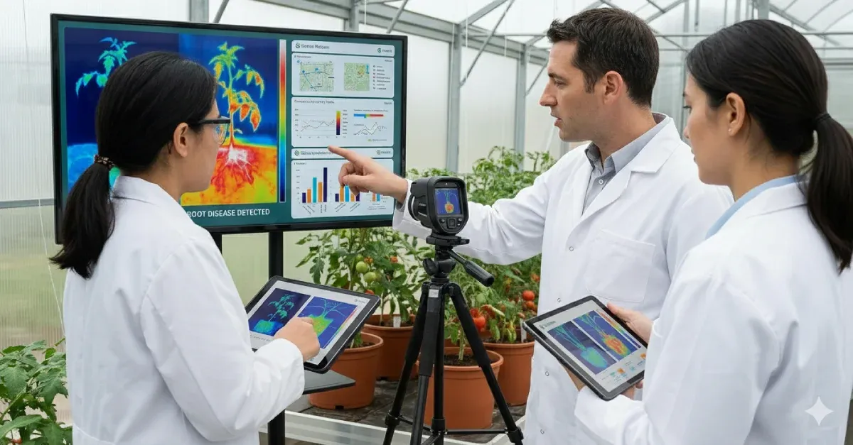
Practical Implementation: Step-by-Step Thermal Imaging Workflow
If we want to implement thermal imaging plractically we require a structured workflow that spans equipment selection, image acquisition, data preprocessing, and standardization of measurement conditions. Each step plays a critical role in ensuring thermal data accurately reflect plant physiological status and can be reliably used for downstream analysis and machine learning-based classification. The following subsections outline key methodological considerations reported in recent thermal imaging and plant phenotyping studies.
Equipment Selection
Thermal cameras used in agricultural and plant phenotyping applications typically operate in the long-wave infrared (LWIR) region, where vegetation exhibits high and relatively stable emissivity. Sensor selection should prioritize adequate spatial resolution to capture temperature heterogeneity across plant surfaces, as well as high thermal sensitivity to detect small temperature differences associated with early stress responses. Radiometric capability and proper emissivity configuration are essential to ensure that recorded thermal values correspond to actual surface temperatures rather than relative intensity measures.
Image Acquisition Protocols
Accurate thermal image acquisition depends on consistent imaging protocols, including fixed spatial resolution, appropriate frame rates, and careful radiometric calibration. Calibration procedures are necessary to compensate for sensor drift and environmental influences, enabling conversion of recorded radiance into reliable temperature measurements. Acquisition timing, camera-to-canopy distance, and viewing angle should remain consistent throughout data collection to allow meaningful temporal and spatial comparisons.
Image Preprocessing
Raw thermal images often require preprocessing before analysis to reduce noise and isolate plant regions from background elements. Common preprocessing steps include noise filtering, contrast enhancement, and segmentation of plant areas based on thermal or co-registered visual information. Segmentation enables extraction of temperature values specifically from plant tissues, which is essential for accurate feature computation and disease-related analysis.
Data Collection in Greenhouses vs. Field Conditions
Thermal data collection in greenhouse environments benefits from controlled temperature, humidity, and airflow, which reduces environmental variability and improves measurement repeatability. In contrast, field-based thermal imaging is influenced by fluctuating environmental conditions such as wind, solar radiation, and ambient temperature gradients. These factors can introduce variability in thermal measurements and must be carefully considered when interpreting temperature-based stress indicators under open-field conditions.
Building Robust Datasets for Machine Learning
Robust machine learning models for thermal disease detection require datasets that capture variability across plant growth stages, disease severity levels, and environmental conditions. Limited sample sizes and narrow data diversity can reduce model generalizability and increase the risk of overfitting. Therefore, repeated measurements and inclusion of diverse experimental conditions are recommended to improve dataset representativeness and model robustness.
Standardizing Measurement Conditions for Consistency
Standardization of measurement conditions is essential for ensuring consistency and comparability of thermal data across experiments. This includes maintaining fixed emissivity settings, consistent camera configuration, and controlled acquisition protocols. Environmental parameters should be monitored and documented, as variations in atmospheric conditions can significantly affect thermal measurements. Standardized workflows support reproducibility and reliable interpretation of temperature-based indicators in plant disease detection studies.
Conclusion
Thermal infrared imaging has emerged as a game-changing technology for plant disease management by enabling non-destructive, continuous, and early detection of physiological stress associated with root diseases. By capturing subtle temperature variations linked to disruptions in water uptake, transpiration, and stomatal regulation, thermal imaging provides a valuable indirect window into belowground disease processes that often remain undetected using conventional diagnostic methods. However, transforming thermal data into actionable insights requires robust analytical frameworks, where machine learning plays a critical role in extracting meaningful patterns from high-dimensional temperature information and enabling automated, scalable disease diagnosis. When integrated with modern precision agriculture platforms, thermal imaging can complement existing sensing technologies and support data-driven decision-making in crop protection and resource management. Looking ahead, the combination of thermal sensing, advanced analytics, and intelligent platforms opens new opportunities for practical deployment at scale, where solutions such as Sairone platform that developed by Saiwa can facilitate the translation of thermal intelligence into operational tools for early disease detection, monitoring, and sustainable agricultural management.
References (4)
Comments
No comments yet!
Table of Contents
No headings were found on this page.